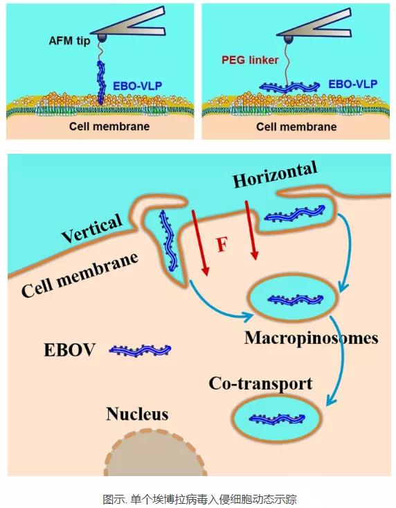
近日,病毒学国家重点实验室崔宗强研究组、长春理工大学单玉萍研究组和国家纳米科学技术中心施兴华研究组合作在《ACS Nano》期刊发表了题为“Entry Dynamics of Single Ebola Virus Revealed by Force Tracing”的文章。研究表明,基于单颗粒力跟踪测试,分子动力学模拟和单颗粒荧光跟踪的发现,提出了纤维EBO-VLP细胞沿平行/垂直方向进入的可能模型。 水平或垂直状态下的EBO-VLP通过巨泡细胞样途径进入细胞。 随后,将EBO-VLP和细胞大脂质体在细胞质中共转运进行感染。 这项工作揭示了单颗粒水平上EBO-VLPs感染的动力学机制,这有助于更好地了解感染。
研究人员首先构建了丝状埃博拉病毒样颗粒(EBO-VLP)并对其进行荧光标记,该丝状结构EBO-VLP和野生型EBOV具有同样的入侵细胞能力,但其内部没有病毒核酸,不能复制。研究人员利用双功能PEG Linker将EBO-VLP连接到原子力显微镜的探针上,通过力示踪技术实时监测单个EBO-VLP内吞进入细胞的动态过程。结果发现EBO-VLP可以通过水平或垂直两种模式进入细胞,两种模式对应的力和时间不同。对两种模式进行分子动力学模拟,也说明EBO-VLP以垂直方向进入细胞比水平模式所需时间更长,所需能量(也就是力)更大。
通过分析计算力示踪检测到的内吞力信号,推测大约有九个受体结合位点在EBO-VLP入侵过程中参与病毒内吞。实时单颗粒荧光示踪显示EBO-VLP与细胞巨胞饮标志物能够很好地共定位,并具有相同的运动速率、轨迹、均方位移等,巨胞饮抑制剂可以显著抑制其入侵,从而可视化地证实了EBO-VLP是以巨胞饮途径入侵细胞。

文章实时动态解析了单个EBO-VLP入侵宿主细胞过程,揭示了丝状EBO-VLP以水平或垂直两种模式进入细胞,以及对应的力学、时-空、能量、与受体作用方式、入侵途径等精细机制,对深入理解EBOV的感染机理具有重要意义,也为开发抗病毒途径提供了基础。(论文链接:https://pubs.acs.org/doi/10.1021/acsnano.0c01739)
ABSTRACT:
Infections by the Ebola virus (EBOV) rapidly cause fatal hemorrhagic fever in humans. Viral entry into host cells is the most critical step in infection and an attractive target for therapeutic intervention. Herein, the invagination behavior and entry dynamics of filamentous Ebola virus-like particles (EBO-VLPs) were investigated using a force tracing technique based on atomic force microscopy and single-particle fluorescence tracking in real time. The filamentous EBOVVLPs might enter cells in both horizontal and vertical modes, and the virus−receptor interactions during endocytic uptake were analyzed. In addition, molecular dynamics simulations and engulfment energy analysis further depicted EBO-VLP entry in the horizontal and vertical directions and suggested that internalization in the vertical direction requires a larger force and more time. This report provides useful information for further revealing the mechanism of viral infection, which is important for understanding viral pathogenesis.
CONCLUSIONS:
Based on the findings from a single-particle force tracing test, molecular dynamics simulation, and single-particle fluorescence tracking, we proposed a possible model of fibrous EBO-VLP cell entry in parallel/vertical directions. EBO-VLPs under horizontal or vertical enter into cells via a macropinocytosis-like pathway. Subsequently, the EBO-VLPs and cellular macropinosomes are cotransported in the cytoplasm for infections. This work reveals the dynamic mechanism of EBO-VLPs infection at the singleparticle level, which is helpful for better understanding infections by the Filoviridae family.
转载自:病毒学界